Much of our efforts are driven by the hypothesis that the vast majority of orthopaedic shoulder disorders are related to dysfunction of the rotator cuff muscles and tendons. These intrinsic shoulder muscles play a large role in helping to stabilize the shoulder and injury to them can result in common shoulder pathologies, such as impingement syndrome, rotator cuff tears, instability, occupational disorders and possibly osteoarthritis. The clinical occurrence of shoulder pain is second only to back and neck pain and for just occupational disorders, there is an annual financial impact in this country approaching $6 billion due to utilization of healthcare services, lost work days and worker disability costs.
A comprehensive review by the National Institute for Occupational Safety and Health has found compelling evidence for a “positive association between highly repetitive work and shoulder musculoskeletal disorders.” While it is possible that repetitive arm motion can theoretically lead to direct tendon damage due to overload, the flow chart below demonstrates an alternative model linking repetitive arm motion and injury.
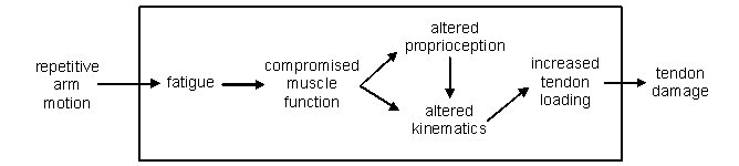
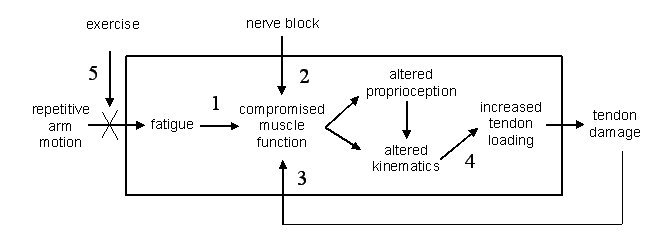
We hypothesize that repetitive arm motion resulting in compromised muscle function due to fatigue leads to altered proprioception and kinematics, which could result in an increase in tendon loading and ultimately tendon damage. Given this model, we have identified several key links in the pathway where we are focusing our research efforts. These include the effects of 1) fatigue, 2) nerve blocks, 3) tendon damage, 4) altered kinematic, and 5) exercise:
1) Muscle Fatigue
We have completed two studies examining the effects of different fatigue protocols on scapular kinematics. One fatigue protocol specifically targeted the rotator cuff and one was a more general fatigue protocol. A similar pattern of scapular kinematic changes was noted with these two very different fatigue studies. Additionally, electromyographic data indicated that one of the rotator cuff muscles (infraspinatus) demonstrated the most local fatigue in both protocols. These results suggests that rotator cuff muscle fatigue may play a significant role in altering scapular kinematics and there may be a common pattern of kinematic changes secondary to shoulder muscle fatigue regardless of how fatigue is induced. It is interesting to note that the findings related to scapular kinematics are similar to those in studies that have investigated scapulothoracic motion in subjects with rotator cuff tears. This suggests that there may be an inherent compensatory mechanism whereby scapular motion is altered in response to impairments of the rotator cuff muscles. In conjunction with Dr Laurel Kincl at the Labor Education and Research Center on campus, we have designed a field based study of the effects of fatigue on shoulder kinematics and proprioception due to a typical workday in dental hygienists, which was recented funded by NIOSH. This would be the first study to examine the effects of repetitive shoulder motion on kinematics and proprioception in an actual workplace setting and represents the next step towards understanding the progression from repetitive arm elevation to increased tissue loading. The results may also serve as a basis for selecting effective exercise strategies aimed at reducing fatigue and improving endurance for occupational tasks.
2) Suprascapular Nerve Block
In collaboration with Dr Peter Kosek, a local anesthesiologist specializing in pain management, we have conducted a study of the effects of a pharmacological suprascapular nerve block on scapular kinematics. This nerve was selected since this it innervates the two rotator cuff muscles most affected by cuff pathologies (supraspinatus and infraspinatus). Despite the fact that the muscles innervated by the suprascapular nerve do not directly control the movement of the scapula, they appear to result in a compensatory increase in scapular motion. We are planning on performing a follow-up study to examine the effects of this block on translations at the glenohumeral joint with the use of fluoroscopy (digital x-rays). Since an increase in superior translation is directly related to increased loading of the rotator cuff tendons, this study will further our understanding of the relationship between rotator cuff dysfunction and tendon loading. This project was also funded on the same NIOSH grant as with the dental hygienists. We are currently using a cadaver model to validate this fluoroscopic method.
3) Rotator Cuff Tear
We am currently working on a grant from the Oregon Medical Research Foundation to compare scapular kinematics in patients with full thickness rotator cuff tears and healthy control. This work is in collaboration with orthopaedic surgeons from Orthopaedic Healthcare Northwest. Although this work is still in progress, it is anticipated that patients with rotator cuff tears will demonstrate more scapular motion during active arm elevation when compared to healthy controls. However, one of the main mechanistic questions associated with this phenomena relates to whether this is a biomechanical mechanism (ie, the glenohumeral joint is tight, so in order to achieve the same elevation angle, more motion needs to come from the scapula) or if it is a motor control mechanism (ie, the glenohumeral joint has a normal range of motion, it is just not being utilized). That is why we are also assessing passive shoulder motion, which ideally removes the motor control part of the equation. If the passive motion is the same for the two groups, we would conclude that it is more of a motor control problem. However, if the passive motion pattern mirrors the active motion pattern (more scapulothoracic motion for the rotator cuff group), then we would conclude that it is a biomechanical problem. Either way, this study will not only describe kinematics patterns, it will help explain them.
4) Cadaver Model
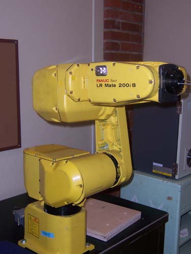
Although measurements of in-vivo kinematics are important, we also need to examine the effects of kinematic alterations on joint contact forces. We have developed a cadaver model to examine the effects of scapular orientation on clearance in the subacromial space (this is the area where the rotator cuff tendons are located). Human cadaver joints were secured to a mechanical testing machine and forces were applied to simulate muscular contractions. Translations of the glenohumeral joint were simulated and the distance before the development of significant contact force was measured. Specimens were testing at varying scapular angles and subacromial clearance was found to decrease with an increase in scapular rotation. These results would suggest that the increases in scapular rotations noted in three reported models of rotator cuff dysfunction (fatigue, nerve block and rotator cuff tears) may represent a positive compensation that serves to open up the subacromial space and reduce contact forces. The lab has purchased a 6 DOF robotic manipulator that will be used to further refine this model.
5) Exercise
In the future, we plan on evaluating the effectiveness of a training program on reducing fatigue-induced proprioceptive deficits caused by repetitive arm motion. It is possible that an endurance exercise program will reduce proprioceptive deficits by increasing the resistance of the shoulder muscles to fatigue. This study would represent a step towards understanding how to disrupt the progression from repetitive arm elevation to abnormal joint loading. |